What Is Autophagy? The Nobel Prize Biology
In 2016, Japanese cell biologist Yoshinori Ohsumi received the Nobel Prize in Physiology or Medicine for his work decoding the molecular machinery of autophagy — a word derived from the Greek for "self-eating." Ohsumi's group used budding yeast (Saccharomyces cerevisiae) in the early 1990s to identify the core autophagy genes (ATG genes), demonstrating that cells have an ancient, conserved system for digesting their own components. That system, it turns out, is one of the most important quality-control mechanisms in biology.
Autophagy is the process by which cells engulf damaged proteins, misfolded aggregates, dysfunctional organelles, and intracellular pathogens within double-membrane vesicles called autophagosomes, which then fuse with lysosomes to be digested and recycled. The resulting amino acids, lipids, and nucleotides re-enter the cytoplasmic pool to be reused for energy production or biosynthesis. This is not a passive recycling bin — it is a precisely regulated, energy-sensitive quality-control system that responds dynamically to nutrient status, stress, and cellular damage.
There are three primary forms: macroautophagy (the bulk process typically referred to simply as "autophagy"), microautophagy (direct lysosomal invagination), and chaperone-mediated autophagy (CMA), which targets specific proteins bearing a KFERQ-like pentapeptide motif. In the context of fasting and health, macroautophagy is the dominant form and the one most thoroughly studied in relation to nutrient deprivation.
The Molecular Switch: mTOR and AMPK
Autophagy is controlled by two opposing kinase systems that together act as the cell's nutritional sensor. Understanding them is essential to understanding the fasting timeline.
mTORC1 (mechanistic target of rapamycin complex 1) is the master brake on autophagy. When nutrients — particularly amino acids and glucose — are abundant, mTORC1 is active. It phosphorylates and inactivates the ULK1/2 complex, the initiator of autophagosome formation. Active mTORC1 essentially keeps autophagy in a suppressed, low-activity state. Insulin signaling also activates mTORC1 via the PI3K/Akt/TSC1/2 axis, which is why postprandial states (high insulin) are characterised by low autophagy.
AMPK (AMP-activated protein kinase) is the reciprocal sensor — it is activated when the AMP:ATP ratio rises, indicating cellular energy deficit. AMPK activates autophagy via two routes: (1) direct phosphorylation of ULK1 at activating sites (Ser317, Ser777), and (2) phosphorylation of Raptor (part of mTORC1), which inhibits mTOR activity. AMPK activation is the central signal linking fasting, caloric restriction, and exercise to autophagy induction.
A third layer involves the transcription factor TFEB (transcription factor EB), which is normally sequestered in the cytoplasm by mTORC1-mediated phosphorylation. When mTORC1 is suppressed during fasting, TFEB translocates to the nucleus and transcriptionally upregulates both autophagy genes and lysosome biogenesis genes — amplifying autophagic capacity over hours to days.
The Fasting Timeline: Hour by Hour
The progression from fed to deeply autophagic state is not a single threshold event but a gradual continuum shaped by glycogen stores, metabolic health, and prior meal composition.
| Fasting Duration | Primary Biochemical Event | Autophagy Status |
|---|---|---|
| 0–4 hours | Fed/postprandial: high insulin, active mTORC1 | Basal (suppressed) |
| 4–8 hours | Insulin declining, glycogen still ample | Basal rising slightly |
| 8–12 hours | Hepatic glycogen ~50% depleted, glucagon rising | Modest upregulation beginning |
| 12–16 hours | Glycogen near-depleted, ketogenesis starting, AMPK activating | Significant upregulation; mTORC1 suppression ongoing |
| 16–24 hours | Ketones rising, gluconeogenesis dominant, mTORC1 substantially suppressed | Peak flux in liver; robust upregulation in most tissues |
| 24–48 hours | Deep ketosis, systemic AMPK activation, TFEB nuclear | Near-maximal; mitophagy prominent |
| 48–72 hours | Protein conservation mechanisms active, growth hormone elevated | Maximal recycling of long-lived proteins and organelles |
The 12-hour glycogen depletion point is the gateway. The liver stores roughly 80–100 grams of glycogen under typical conditions; skeletal muscle holds an additional 300–400 grams but cannot export glucose to the circulation. Once hepatic glycogen is depleted, the liver switches to gluconeogenesis and ketogenesis, insulin falls to near-basal levels, and the conditions for mTOR suppression are established. This is why the overnight fast (12–14 hours from dinner to breakfast) is the minimum window at which meaningful autophagy upregulation can begin — though it does not reliably produce the peak flux seen at 18–24 hours.
Individual Variation: Why the Timeline Differs Between People
The fasting timeline described above represents an average in metabolically healthy individuals. Several factors shift it significantly.
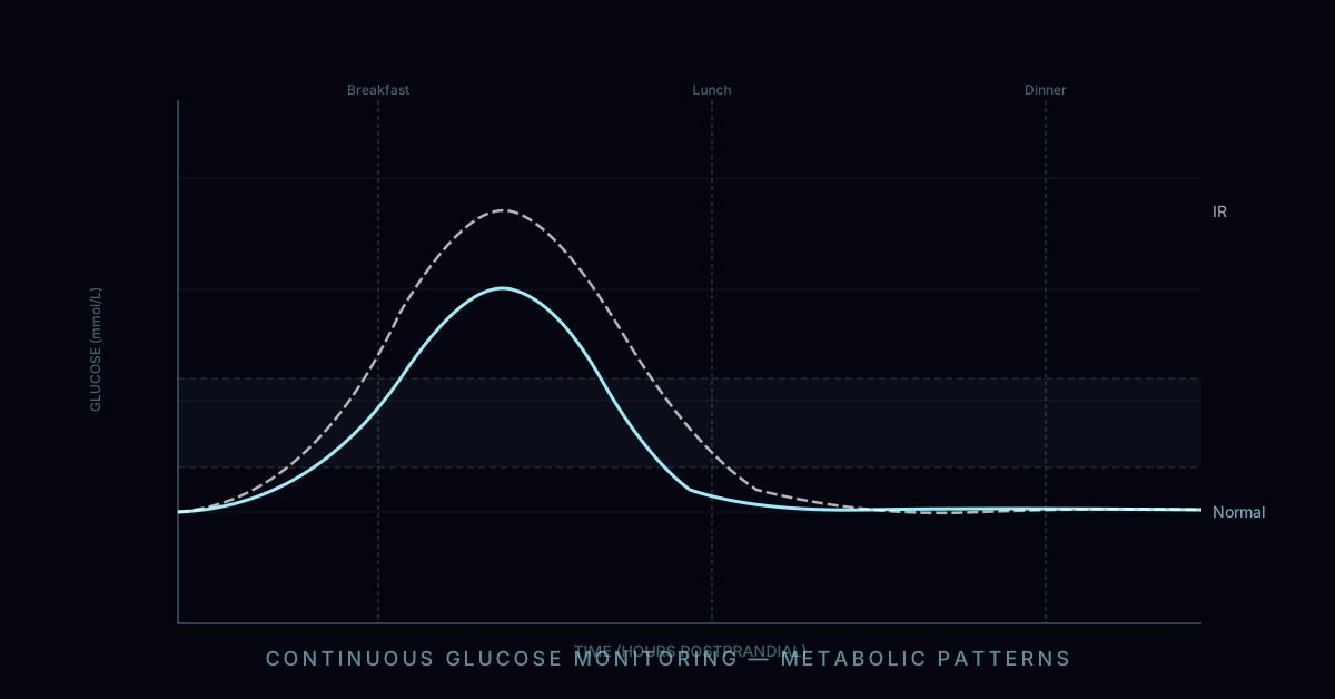
Insulin resistance and metabolic syndrome delay autophagy induction substantially. In insulin-resistant individuals, basal insulin is chronically elevated, mTORC1 remains partially active even between meals, and hepatic glycogen stores may be partially replenished by gluconeogenesis even during short fasts. Studies examining LC3-II/p62 markers in people with type 2 diabetes versus controls consistently show blunted autophagic response to equivalent fasting durations. These individuals may need 20–24 hours before achieving autophagic flux equivalent to what a metabolically healthy person achieves at 16 hours.
Prior meal composition matters enormously. A high-carbohydrate meal (pasta, rice, bread) consumed the evening before a fast maximally fills hepatic glycogen and triggers a large insulin response — autophagy induction will begin later than if the last meal was low-carbohydrate or protein-dominant. Conversely, someone who consumed a low-carbohydrate, moderate-protein dinner may have hepatic glycogen substantially depleted by 8–10 hours post-meal.
Exercise history and glycogen status: an individual who exercised vigorously (depleting muscle glycogen) before beginning a fast will reach the conditions for autophagy induction sooner, since total body glycogen load is lower. This is one mechanism underlying the additive benefit of exercising in a fasted state.
Age and lean mass: older adults with reduced muscle mass have smaller total glycogen stores, which in principle could accelerate the glycogen depletion phase — though age-related impairments in AMPK sensitivity and mitochondrial function may partially offset this.
How Autophagy Is Measured (And Why There Is No Consumer Test)
The definitive measurement of autophagic flux in live cells uses genetically encoded fluorescent reporters — typically a tandem mCherry-GFP-LC3 construct, which appears yellow in autophagosomes (both fluorophores active) and red in autolysosomes (GFP quenched by the acid environment), allowing real-time quantification of flux rather than just static LC3-II accumulation. This requires transfected cell lines or transgenic animals — it is not applicable to clinical samples.
In human research, the most used proxy is p62/SQSTM1, a scaffold protein that is specifically recognised and degraded by autophagy. Falling p62 over a fasting time course indicates increasing autophagic flux. Conversely, accumulated p62 is a hallmark of autophagy impairment in disease (e.g. in the hepatocytes of people with non-alcoholic fatty liver disease). LC3-II protein levels in peripheral blood mononuclear cells (PBMCs) obtained from blood draws have also been used as a proxy, though interpretation requires understanding that LC3-II accumulates both when autophagy is induced and when lysosomal clearance is blocked.
Several consumer products claim to measure autophagy — typically via blood, urine, or breath tests. None has been independently validated against gold-standard flux assays. The most scientifically grounded consumer approach remains using fasting duration as the proxy, combined with metabolic state markers such as blood ketone levels (beta-hydroxybutyrate >0.5 mmol/L indicates established ketosis, which co-occurs with robust autophagy induction).
Exercise and Autophagy: AMPK as the Common Thread
Exercise is an independent autophagy inducer that acts via the same AMPK pathway as fasting. During moderate-to-vigorous aerobic exercise, ATP turnover rates in active muscle increase 100-fold over rest, sharply raising the AMP:ATP ratio and activating AMPK. A landmark 2012 paper by He et al. published in Nature used mice expressing an autophagy-deficient form of Bcl-2 to demonstrate that exercise-induced autophagy is not merely a correlate of exercise but is required for its metabolic benefits — autophagy-blocked mice could not maintain blood glucose or sustain endurance performance.
Human biopsy studies confirm increased LC3-II and reduced p62 in skeletal muscle within 30–60 minutes of acute exercise at >60% VO₂max. The combination of fasting and exercise appears additive: exercising in the fasted state (e.g., morning training after an overnight fast) amplifies AMPK activation and prolongs the window of mTOR suppression compared to fed-state exercise. The practical implication is that people who exercise while in a fasted state are likely achieving greater autophagic stimulus per session, though this must be balanced against reduced performance capacity for high-intensity efforts.
Autophagy in Disease Context: Why It Matters
The health rationale for optimising autophagy extends beyond general wellness. Impaired autophagy has been mechanistically linked to: (1) neurodegeneration — in Parkinson's, Alzheimer's, and Huntington's disease, protein aggregates (alpha-synuclein, tau, polyglutamine) accumulate specifically because autophagy-mediated clearance is insufficient; (2) cancer — autophagy has a complex dual role (tumour suppressive in early carcinogenesis, potentially pro-survival in established tumours); (3) ageing — the rate of autophagic flux declines with age, and pharmacological or dietary enhancement of autophagy extends lifespan in every model organism tested; (4) metabolic disease — lipophagy (selective autophagy of lipid droplets) is a primary mechanism for hepatic lipid clearance, and its impairment contributes to non-alcoholic fatty liver disease.
Rapamycin, the mTOR inhibitor used in organ transplantation, is the most studied pharmacological autophagy inducer — it extends lifespan in mice even when administered late in life, and is currently being investigated in human ageing trials. The relevance to fasting is that both interventions converge on the same mTORC1 axis.