Quick Answer
A blood sugar spike (postprandial hyperglycaemia) is a rapid rise in blood glucose after eating, typically above 140 mg/dL (7.8 mmol/L) at one hour, followed by a rapid insulin-driven fall. Symptoms include fatigue, brain fog, renewed hunger, irritability, and blurred vision. Spikes cause oxidative stress, endothelial damage, and glycation even in people without diabetes. The most effective prevention strategies are meal sequencing (vegetables and protein before carbohydrates), post-meal walking, and avoiding refined carbohydrates and liquid sugars.
What a Blood Sugar Spike Actually Is
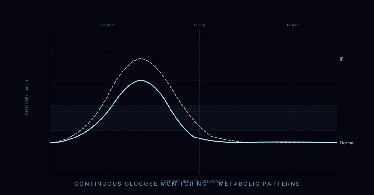
After you eat carbohydrates, your digestive system breaks them down into glucose, which enters the bloodstream via the small intestine. Blood glucose rises, the pancreatic beta cells sense this rise and secrete insulin, and insulin signals cells throughout the body — primarily muscle, liver, and adipose tissue — to take up glucose from the blood. This process returns blood glucose toward the fasting baseline, typically within 2–3 hours in people with healthy glucose metabolism.
A blood sugar spike refers to the abnormally rapid and high excursion from baseline that occurs when this system is overwhelmed — either because too much glucose was delivered too quickly (a large serving of quickly-digested carbohydrates), because insulin secretion or sensitivity is impaired, or both. The clinical threshold most commonly used is a postprandial glucose above 140 mg/dL (7.8 mmol/L) at one or two hours after eating, which is the diagnostic cut-off for impaired glucose tolerance in a 2-hour oral glucose tolerance test. However, research using continuous glucose monitoring (CGM) shows that the damage begins below this threshold: even excursions to 120–140 mg/dL produce measurable increases in oxidative stress markers and endothelial dysfunction.
The size and frequency of postprandial spikes are now understood to be important independent predictors of cardiovascular disease, cognitive decline, and all-cause mortality, separate from and additive to fasting glucose and HbA1c. This matters because standard diabetes screening — which measures fasting glucose and HbA1c — can miss the people who spike largest after meals while maintaining normal fasting levels. The DECODE study, which tracked 25,000 people across Europe, found that postprandial glucose was a stronger predictor of cardiovascular mortality than fasting glucose, and that many of those who died had never received a diabetes diagnosis.
Symptoms of a Blood Sugar Spike
The symptoms of a blood sugar spike follow a predictable temporal pattern tied to the glucose-insulin curve. Understanding this timeline helps you recognise what your body is telling you after meals.
| Phase | Timing | What's happening / Symptoms |
|---|---|---|
| Rapid rise | 0–60 min | Glucose floods the bloodstream. Brief energy surge, increased alertness. Some people notice warmth or flushing. Blood is thickened by osmotic load — some people notice visual blurring as the lens absorbs fluid. |
| Insulin peak | 60–90 min | Large insulin secretion rapidly clears glucose. Fatigue and heaviness begin. Brain fog emerges as glucose falls sharply. Yawning. Heart palpitations in some people (from adrenaline secreted in response to falling glucose). |
| Crash / trough | 90–180 min | Reactive hypoglycaemia if insulin overshoot occurs. Intense hunger and carbohydrate cravings despite having recently eaten. Irritability, difficulty concentrating, shakiness, anxiety, cold sweat. Strong urge to snack. |
| Recovery | 180–240 min | Counter-regulatory hormones (glucagon, cortisol, adrenaline) raise glucose back toward baseline. Symptoms resolve but baseline cortisol is elevated — adding to stress load across the day. |
Not all spikes produce all symptoms. People who eat high-carbohydrate diets consistently tend to adapt — their brains become accustomed to running on high-glucose fuel and have reduced sensitivity to the acute signals. This adaptation is not protective; it means the metabolic damage continues without the warning system. People transitioning from low-carbohydrate eating to high-carbohydrate eating, or vice versa, tend to feel symptoms most acutely because their physiology is less adapted.
The most clinically underrecognised symptoms of chronic blood sugar volatility — as opposed to acute individual spikes — are the cumulative effects on mood and cognition: persistent mid-afternoon energy crashes, difficulty sustaining focus for more than 45–60 minutes, irritability in the pre-meal period, and the subjective sense of being mentally dulled or slow in the hours after high-carbohydrate meals. These symptoms are so normalised in industrial-diet populations that most people assume they are intrinsic to how they function, rather than recognising them as the response of a glucose-dependent brain to metabolic instability.
The Physiology: Why Spikes Happen
The glucose-insulin axis is a tightly regulated feedback system with multiple control points. Understanding where it can go wrong explains both the causes of spikes and the mechanisms behind the most effective interventions.
Gastric Emptying Rate
The first control point is how fast the stomach delivers food to the small intestine. Gastric emptying rate is a primary determinant of how quickly glucose enters the bloodstream. Fat, protein, fibre, and acids (vinegar, lemon juice) in a meal all slow gastric emptying, spreading the glucose load over a longer period and producing a shallower, broader rise rather than a sharp spike. Liquid meals — particularly sugary drinks — bypass this control entirely because they require no mechanical digestion and empty from the stomach very rapidly. This is why liquid calories from sodas, fruit juice, and sweetened beverages are uniquely spiking: their glucose reaches the small intestine in minutes rather than the 1–2 hours that a solid mixed meal takes.
Small Intestinal Absorption
Once in the small intestine, carbohydrates are absorbed via sodium-glucose co-transporters (SGLT1) and glucose transporters (GLUT2). The rate of absorption is proportional to the glucose concentration gradient — which is why a very large carbohydrate load, even if delivered slowly, can still produce a steep rise. Dietary fibre slows this absorption by increasing intestinal viscosity and forming a physical barrier. This is why intact whole grains and legumes produce substantially smaller glucose excursions than the same calories from refined flour or white rice: the fibre matrix slows both gastric emptying and intestinal absorption.
Insulin Secretion: First Phase and Second Phase
Pancreatic beta cells secrete insulin in two phases. The first-phase response is a rapid burst of pre-formed insulin within 1–5 minutes of eating — triggered not by glucose itself but by the cephalic phase (anticipatory signals from smell, taste, and meal initiation) and incretin hormones (GLP-1 and GIP) released from the gut. This early insulin release is critical: it suppresses hepatic glucose output and prepares peripheral cells to receive glucose before the postprandial wave arrives. In people with impaired first-phase insulin secretion — which is one of the earliest detectable defects in type 2 diabetes, often visible years before fasting glucose abnormalities develop — the early suppression of hepatic glucose output fails, and postprandial spikes are much larger and more prolonged. The second-phase insulin response follows 10–60 minutes later, driven by the rising blood glucose itself.
Peripheral Insulin Sensitivity
Even with normal insulin secretion, blood glucose will spike higher and remain elevated longer if the target tissues — primarily skeletal muscle — are insulin resistant and require more insulin to achieve the same glucose uptake. Skeletal muscle normally accounts for 70–80% of postprandial glucose disposal. Insulin resistance in muscle is driven by intramyocellular lipid accumulation (particularly diacylglycerol and ceramide species derived from saturated fat and obesity), physical inactivity (GLUT4 transporter density falls rapidly with disuse), and chronic inflammation. This is the vicious cycle: high glucose spikes drive insulin resistance, and insulin resistance drives higher glucose spikes.
Why Spikes Are Harmful: The Damage Mechanisms
The harm from blood sugar spikes is not merely the peak glucose value — it is the acute biological events that the rapid rise triggers. Three mechanisms are particularly well-documented:
1. Oxidative Stress Burst
When mitochondria are overwhelmed by excess glucose substrate, they generate a burst of reactive oxygen species (ROS) — superoxide, hydrogen peroxide, hydroxyl radical — that damage mitochondrial DNA, membrane lipids, and proteins. Work by Antonio Ceriello and colleagues demonstrated that this oxidative burst is proportional to the size of the glucose excursion, not the absolute glucose level, and that it can be measured in normal-weight healthy adults after a single high-glycaemic meal. Antioxidant supplementation does not prevent it because the ROS originate inside mitochondria, where orally-absorbed antioxidants do not reach in effective concentrations. The only effective suppression is preventing the spike itself.
2. Endothelial Dysfunction
The endothelium — the single-cell layer lining every blood vessel — becomes temporarily dysfunctional during a glucose spike. Nitric oxide (NO) production falls (reducing vasodilation and blood flow), adhesion molecules are upregulated (promoting monocyte adhesion — the first step in atherosclerotic plaque formation), and von Willebrand factor is released (promoting clotting). These effects are measurable within 1–2 hours of a high-glycaemic meal and resolve as glucose returns to baseline — but repeat exposure across decades accumulates damage. Population studies show a strong dose-response between postprandial glucose excursion magnitude and carotid intima-media thickness, a marker of early atherosclerosis.
3. Advanced Glycation End-products (AGEs)
Glycation is the non-enzymatic bonding of glucose to proteins and lipids, forming advanced glycation end-products (AGEs). This process is irreversible and accumulates with cumulative glucose exposure — both the peak and the duration of elevation matter. AGEs cross-link collagen (stiffening arterial walls, skin, and the lens of the eye), modify LDL particles (making them more pro-atherogenic), and activate the RAGE receptor on endothelial cells and macrophages (driving chronic inflammation). HbA1c — the standard diabetes monitoring test — is itself a glycation product of haemoglobin; its 90-day averaging reflects cumulative glycation exposure, but misses the large excursions that drive disproportionate AGE formation.
What CGM Data Reveals: Individual Variability
One of the most important findings from consumer CGM research — particularly from studies at the Weizmann Institute and from large citizen-science datasets — is the extraordinary degree of individual variation in postprandial glucose responses. Two people can eat identical meals and produce completely different glucose curves. The same person can produce different responses to the same meal depending on the time of day eaten, the preceding night's sleep quality, stress levels, the previous day's exercise, and gut microbiome composition.
Key CGM findings in non-diabetic populations
This variability is why generic glycaemic index tables are an imperfect guide. Bread may spike one person to 160 mg/dL and another to 115 mg/dL. The only way to know your personal response is to measure it. A two-week CGM trial — now accessible without prescription via devices like the Lingo, Stelo, or Libre Sense — provides more personalised metabolic insight than any population-average glycaemic index table. Wearing one while eating your normal diet for one week, then experimenting with meal sequencing and post-meal walks in the second week, typically produces both insight and motivation that are difficult to obtain any other way.
Foods That Spike and Foods That Don't
The glycaemic response to a food depends on: its total glucose content, how quickly it is digested and absorbed (glycaemic index), the total amount eaten (glycaemic load), and what it is eaten with. Here is a practical orientation:
| Spike level | Foods | Reason |
|---|---|---|
| Very high | Sodas, fruit juice, sports drinks, glucose tablets, white bread, white rice, instant oats, rice cakes, most breakfast cereals, baked potato, candy, syrup | Fast-digesting, high glucose density, no buffering fibre or fat |
| Moderate | Whole fruit (not juice), rolled oats, pasta, sourdough bread, brown rice, sweet potato, most dried fruit, dark chocolate, honey | Higher fibre, lower processing, but still significant glucose content |
| Low | All non-starchy vegetables, most legumes (lentils, chickpeas, black beans), nuts, seeds, most whole dairy, eggs, meat, fish, avocado, olive oil | Very low glucose content, or slow digestion with high fibre/fat/protein |
| Context-dependent | White rice with lots of fat and fibre; bread eaten after a large salad; any carbohydrate eaten after a protein/fat starter; same meal eaten at 8 AM vs. 8 PM | Meal composition and timing modify the response dramatically |
A critical practical point: fructose does not directly spike blood glucose — it is metabolised almost entirely by the liver. This is why agave syrup and high-fructose corn syrup can score low on blood glucose tests while being metabolically harmful through a different mechanism (excessive hepatic de novo lipogenesis, VLDL secretion, and visceral fat accumulation). Blood sugar tests measure glucose, not metabolic health; fructose overload causes fat liver disease and dyslipidaemia without necessarily raising postprandial glucose. This is one reason why a glucose-only metric misses part of the picture.
How to Prevent Blood Sugar Spikes: Evidence-Based Strategies
Every intervention below has been tested in controlled trials and produces a measurable reduction in postprandial glucose excursion. They are listed roughly in order of evidence quality and practical effect size.
Meal sequencing: vegetables and protein first, carbohydrates last
Multiple RCTs by Alpana Shukla's group at Weill Cornell show that eating fibre and protein before carbohydrates reduces postprandial glucose area-under-the-curve by 30–40% vs. carbohydrates first. The effect is mediated by slowed gastric emptying, blunted first-intestinal glucose absorption, and enhanced GLP-1 secretion from gut L-cells primed by prior protein/fibre contact. Practically: start meals with a salad or vegetables, then protein, then eat carbohydrates. The total food consumed is unchanged; only the order changes. The effect is robust enough to convert a moderate spike into a negligible one for most people.
Post-meal walking (10–20 minutes within 30 minutes of eating)
Skeletal muscle contains the body's largest reservoir of glucose uptake capacity. During exercise, GLUT4 transporters are translocated to the cell surface independently of insulin — meaning muscle can absorb glucose directly without waiting for the insulin response. A 2023 meta-analysis in Sports Medicine found that even light walking (3–5 km/h) within 30 minutes of a meal reduces postprandial glucose by 20–35% compared to seated rest. The effect is specific to the timing: a walk 2 hours after eating has much smaller benefit because the postprandial glucose peak has already passed. The window is 15–30 minutes post-meal for maximum effect.
Vinegar before or during meals (1–2 tablespoons)
Acetic acid (the active component of vinegar) reduces postprandial glucose by approximately 20% through two mechanisms: it inhibits salivary and pancreatic amylase activity (slowing starch digestion) and it slows gastric emptying. The effect is consistent across multiple RCTs and works with apple cider vinegar, white wine vinegar, or any acetic acid source. Diluted in water or as a salad dressing, 1–2 tablespoons before a carbohydrate-containing meal is sufficient. The effect is smaller than meal sequencing but fully additive — both strategies together provide near-complete protection against large spikes from moderate-glycaemic meals.
Replace refined carbohydrates with whole food sources
White bread to sourdough or whole grain reduces postprandial glucose by 20–40% for the same carbohydrate mass. White rice to basmati or parboiled rice (lower amylopectin:amylose ratio) reduces it by 30–50%. Fruit juice to whole fruit eliminates the liquid speed advantage and adds 4–7g of fibre per serving. Each substitution works through the same mechanism: slowing digestion and absorption via intact fibre and cell wall structures. This is not about eating fewer carbohydrates — it is about choosing carbohydrates that arrive more slowly.
Optimise sleep and manage stress
One night of partial sleep deprivation doubles postprandial glucose response the following day. Chronic psychological stress raises cortisol, which directly drives hepatic glucose output and peripheral insulin resistance. These factors are upstream of any dietary intervention — people who consistently sleep 5 hours and work under chronic stress will spike higher than their diet predicts, and no amount of meal sequencing fully compensates. Prioritising sleep to 7–9 hours and implementing a stress management practice (any regular practice with evidence: exercise, meditation, sauna, nature exposure) measurably improves insulin sensitivity within weeks.
Eat carbohydrates earlier in the day
Circadian biology gates insulin sensitivity: peripheral tissues are most insulin-sensitive in the morning and progressively less responsive through the afternoon and evening. The same carbohydrate meal produces a 30–50% larger postprandial glucose excursion when eaten at 6 PM vs. 8 AM. This is why time-restricted eating protocols that concentrate food intake in the morning and afternoon produce metabolic benefits beyond their caloric effects. Practically: make breakfast and lunch the largest carbohydrate-containing meals, and make dinner primarily protein, fat, and vegetables.
When to Seek Medical Evaluation
The strategies above are appropriate for anyone seeking to optimise metabolic health, including people with normal glucose metabolism. However, certain patterns warrant medical evaluation rather than self-management:
Standard fasting glucose and HbA1c testing miss a significant proportion of people with impaired glucose tolerance because they do not capture postprandial excursions. If you have concerning CGM patterns and normal standard labs, an oral glucose tolerance test (OGTT) — which measures glucose at 1 and 2 hours after a 75g glucose load — is the gold-standard test for postprandial glucose metabolism and will detect impairment that fasting tests miss.