What Is Ferritin and Why Does It Matter?
Ferritin is an intracellular iron-storage protein found in virtually every cell of the body. Unlike serum iron — which simply reflects the amount of iron currently circulating in the blood — ferritin represents the body's iron reserve. When iron is absorbed from food or released from recycled red blood cells, excess iron is packaged inside ferritin's protein shell for safe storage, primarily within liver hepatocytes, reticuloendothelial macrophages in the spleen and bone marrow, and skeletal muscle cells. A small amount of ferritin leaks into the bloodstream, and this serum ferritin level serves as the most reliable proxy for total body iron stores in clinical medicine.
Each ferritin molecule can hold up to 4,500 iron atoms within its spherical protein cage — a remarkable design that keeps potentially toxic free iron from generating oxidative damage through the Fenton reaction. Iron in its free form is one of the most potent generators of hydroxyl radicals in biology, which is why the body invests heavily in sequestering it. The flip side of this elegant storage system is that ferritin levels can also rise independently of iron stores when the body is under inflammatory stress, making interpretation more nuanced than a single number suggests.
The serum ferritin test is inexpensive (typically $15 to $40 as a standalone test), widely available, and requires no special preparation beyond fasting for 8 to 12 hours — though even non-fasting ferritin is reasonably informative since it does not fluctuate dramatically with meals the way serum iron does. Despite these advantages, ferritin remains oddly underutilized in routine care, and the reference ranges printed on standard lab reports are so broad as to be nearly meaningless from an optimization standpoint.
The Problem With Standard Reference Ranges
Standard laboratory reference ranges are constructed using a statistical method: collect ferritin values from a large sample of the "normal" population (defined simply as people who are not overtly ill), calculate the 2.5th and 97.5th percentiles, and declare that band the "normal range." This means 95 percent of your sample population will fall within range — including people who are functionally iron-depleted or on the path to iron overload. The range says nothing about optimal function or health outcomes.
The result is a ferritin reference range of 12 to 300 ng/mL for men and 12 to 150 ng/mL for women at most major laboratories. A woman with a ferritin of 14 ng/mL and a woman with a ferritin of 148 ng/mL both receive a "normal" result, yet their physiological states are dramatically different. The first is likely experiencing symptoms of iron deficiency; the second may be perfectly fine or may have inflammatory liver disease — and no distinction is made.
Outcome-based research tells a very different story. Studies examining the ferritin level at which symptoms of iron deficiency resolve, at which endurance performance normalizes, and at which hair cycling returns to normal consistently point to threshold values in the 40 to 70 ng/mL range as the minimum acceptable floor — far above the 12 ng/mL that is technically "within normal." At the upper end, population studies show increasing cardiovascular risk, insulin resistance, and hepatic inflammation above 200 ng/mL in men and 150 ng/mL in women, again well within the standard reference range.
Functional Optimal Ranges: What the Evidence Supports
Based on a synthesis of clinical research and symptom-resolution data, functional medicine practitioners have established the following working optimal ranges for ferritin:
| Population | Standard Reference Range | Functional Optimal Range | Investigation Threshold |
|---|---|---|---|
| Adult men | 12–300 ng/mL | 50–150 ng/mL | >200 ng/mL |
| Premenopausal women | 12–150 ng/mL | 40–100 ng/mL | >150 ng/mL |
| Postmenopausal women | 12–150 ng/mL | 50–150 ng/mL | >200 ng/mL |
| Endurance athletes (all) | 12–300 ng/mL (men) / 12–150 (women) | 80–150 ng/mL | >200 ng/mL |
| Symptomatic investigation floor | — | 30 ng/mL minimum | <30 ng/mL regardless of Hgb |
These ranges are not arbitrary — they correspond to ferritin levels at which tissue iron-dependent enzymes function adequately, at which hair follicle cycling normalizes, at which aerobic capacity is not compromised, and at which the risk of inflammatory or hemochromatotic overload remains low.
Symptoms of Low Ferritin: The Non-Anemic Presentation
One of the most diagnostically overlooked scenarios in primary care is the patient — most often a premenopausal woman or an endurance athlete — who presents with chronic fatigue, hair loss, poor exercise tolerance, restless legs, and cognitive fog, has a normal hemoglobin and hematocrit, and is told their blood work is normal. The missing piece is almost always a ferritin value below 30 ng/mL that was either not measured or dismissed because it was "within range."
Iron is required for the function of hundreds of enzymes across human physiology. Cytochrome c oxidase (Complex IV of the mitochondrial respiratory chain), NADH dehydrogenase (Complex I), and succinate dehydrogenase (Complex II) are all iron-containing enzymes essential for cellular energy production. When ferritin falls below the threshold at which these enzymes are fully supplied, cells shift toward less efficient anaerobic metabolism even at rest, generating the hallmark fatigue that does not respond to rest or sleep.
Hair follicles are among the fastest-proliferating cells in the human body, with each follicle cycling through anagen (growth), catagen (transition), and telogen (resting/shedding) phases every two to six years. Iron deficiency selectively shifts more follicles into the telogen phase simultaneously — the condition known as telogen effluvium — resulting in diffuse scalp hair shedding that typically appears two to four months after the iron-depleting event (heavy periods, pregnancy, surgery, intense dieting). Ferritin levels below 30 ng/mL are consistently associated with telogen effluvium in published studies, and supplementation to raise ferritin above 70 ng/mL is often required before regrowth is observed.
Restless legs syndrome (RLS) — the uncomfortable urge to move the legs, particularly at night — is strongly associated with iron deficiency affecting dopaminergic neurons in the substantia nigra. Iron is required for the activity of tyrosine hydroxylase, the rate-limiting enzyme in dopamine synthesis. Brain iron deficiency can exist even when peripheral ferritin is in the low-normal range, and supplementation to raise ferritin above 50 ng/mL significantly reduces RLS severity in multiple trials.
Other manifestations of functional iron deficiency include: impaired thyroid function (thyroid peroxidase is an iron-heme enzyme required for T4 synthesis and T4-to-T3 conversion); poor cold tolerance; pica (ice eating, or pagophagia, is almost pathognomonic of iron deficiency); worsening of anxiety and attention deficits; and exercise intolerance disproportionate to cardiovascular fitness.
Ferritin as an Inflammation Marker: The High-Ferritin Problem
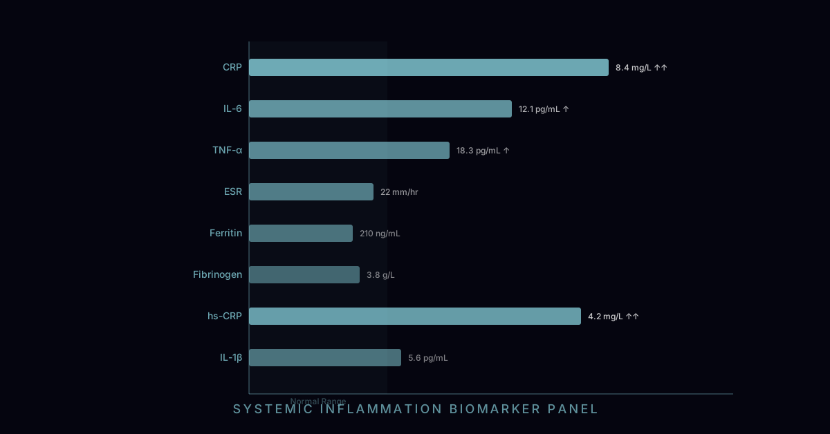
Ferritin is not just an iron-storage protein — it is also an acute-phase reactant, meaning its synthesis is upregulated by interleukin-6 (IL-6), interleukin-1 beta, and TNF-alpha, the same cytokines that drive the inflammatory response. During any significant inflammatory state, ferritin can rise substantially regardless of iron stores, potentially reaching 500, 1,000, or even 10,000 ng/mL in extreme cases (as seen in hemophagocytic lymphohistiocytosis or macrophage activation syndrome).
This creates an important interpretive challenge: elevated ferritin can represent either (1) true iron overload — dangerous iron accumulation damaging organs, or (2) inflammatory elevation — a marker that the body is under significant inflammatory or metabolic stress. Distinguishing these two requires looking at transferrin saturation alongside ferritin. When ferritin is elevated but transferrin saturation is normal or low (below 40%), inflammation is the more likely driver. When both ferritin and transferrin saturation are elevated (saturation above 45%), true iron overload must be investigated.
Elevated ferritin in metabolic syndrome deserves particular attention. Ferritin above 200 ng/mL in a metabolic syndrome patient — even with normal transferrin saturation — has been shown to correlate with hepatic steatosis, liver inflammation, and the degree of insulin resistance. This reflects both the inflammatory nature of metabolic syndrome and the fact that hepatic iron accumulation, even at sub-hemochromatotic levels, worsens insulin signaling and oxidative stress in hepatocytes. The 2019 European Association for the Study of the Liver (EASL) guidelines specifically note that ferritin above 200 ng/mL in women and 300 ng/mL in men with metabolic syndrome should prompt liver assessment regardless of transferrin saturation.
Hereditary Hemochromatosis: The Most Common Genetic Iron Disorder
Hereditary hemochromatosis (HH) is caused by mutations in the HFE gene that impair the body's ability to regulate iron absorption. Normally, the liver protein hepcidin acts as the master regulator of iron, suppressing intestinal iron absorption and macrophage iron release when stores are adequate. HFE mutations impair hepcidin signaling, causing the gut to absorb iron continuously regardless of body stores. Over decades, iron accumulates progressively in the liver, heart, pancreas, joints, skin, and pituitary gland.
The C282Y mutation is responsible for the majority of classic HH cases. Homozygosity for C282Y (inheriting one mutation from each parent) occurs in approximately 1 in 200 individuals of Northern European ancestry, making it one of the most common autosomal recessive disorders in this population. However, clinical penetrance — the proportion of C282Y homozygotes who develop organ damage — is lower than classic genetics would suggest, approximately 28 percent for men and 1 percent for women, because premenopausal menstruation provides continuous iron depletion that delays accumulation.
Untreated hemochromatosis causes liver cirrhosis and hepatocellular carcinoma (the liver absorbs the greatest iron burden), restrictive cardiomyopathy and arrhythmias (cardiac iron deposition), diabetes mellitus (pancreatic beta-cell destruction), arthropathy (iron deposition in chondrocytes, classically affecting the second and third metacarpophalangeal joints — the "iron handshake"), bronze skin discoloration (iron + melanin interaction), and hypogonadism (pituitary iron deposition impairs LH and FSH secretion). These consequences are entirely preventable with early diagnosis and treatment — making the case for proactive ferritin and transferrin saturation testing in at-risk populations.
Consequences of Iron Overload Beyond Hemochromatosis
Even without the HFE mutation, progressive iron accumulation can occur in conditions including thalassemia major (requiring chronic transfusions), alcoholic liver disease (alcohol impairs hepcidin expression), chronic hepatitis C (liver inflammation increases iron deposition), and dysmetabolic iron overload syndrome (DIOS) — a combination of metabolic syndrome and mild-to-moderate iron excess that affects a substantial minority of metabolic syndrome patients.
Iron's toxicity in overload states is mechanistically tied to free radical generation. Excess intracellular iron catalyzes the Fenton reaction (Fe²⁺ + H₂O₂ → Fe³⁺ + OH⁻ + •OH), producing highly reactive hydroxyl radicals that damage DNA, lipid membranes, and proteins. In the liver, this manifests as hepatocyte death and stellate cell activation, driving fibrosis. In the heart, iron accumulation induces cardiomyocyte apoptosis and interstitial fibrosis. In joint cartilage, iron promotes synovial inflammation and accelerates chondrocyte destruction. Recognizing the difference between a ferritin of 180 ng/mL from metabolic inflammation versus 180 ng/mL from early hemochromatosis changes the clinical response dramatically.
The Full Iron Panel: Understanding All Four Markers
Ferritin alone provides an incomplete picture of iron status. A full iron panel including serum iron, TIBC, and transferrin saturation should be ordered whenever ferritin is outside the optimal range or when clinical symptoms suggest iron dysregulation.
Serum iron measures the iron currently bound to transferrin in circulation. It fluctuates significantly throughout the day (highest in the morning, lowest in the evening), with meals, and with stress, making it an unreliable standalone marker. Optimal fasting range is typically 60 to 170 mcg/dL.
Total iron-binding capacity (TIBC) measures the maximum amount of iron that transferrin proteins in the blood can carry. High TIBC (above 400 mcg/dL) indicates the body is producing more transferrin to capture every available iron molecule — a compensatory response to iron deficiency. Low TIBC (below 250 mcg/dL) suggests either iron overload (transferrin is saturated and downregulated) or chronic inflammatory state (inflammation suppresses transferrin production).
Transferrin saturation is calculated as (serum iron ÷ TIBC) × 100. Below 20% with low ferritin = iron-deficiency state. Below 16% = iron-deficient erythropoiesis (the bone marrow does not have enough iron for red blood cell production even if anemia hasn't developed yet). Above 45% = potential iron overload, particularly meaningful if accompanied by elevated ferritin.
Soluble transferrin receptor (sTfR) is a newer marker that reflects the demand for iron at the cellular level. Unlike ferritin, sTfR does not rise with inflammation — it rises specifically with iron deficiency. This makes it valuable for distinguishing true iron-deficiency anemia from anemia of chronic disease, where ferritin is elevated (due to inflammation) but cells are functionally iron-deficient. sTfR levels above 28.1 nmol/L are considered elevated.
Dietary Iron: Heme vs. Non-Heme, Absorption Enhancers and Inhibitors
Not all dietary iron is created equal. Heme iron, found exclusively in animal products (red meat, poultry, fish, shellfish), is absorbed through a dedicated heme-iron transporter (HCP1) at an efficiency of 15 to 35 percent, largely independent of other dietary factors. Non-heme iron, found in plant foods (legumes, spinach, fortified grains, tofu), is absorbed through the divalent metal transporter 1 (DMT1) at an efficiency of only 2 to 20 percent, and this absorption is strongly influenced by other foods consumed at the same meal.
Key absorption enhancers include vitamin C (ascorbic acid), which reduces ferric iron (Fe³⁺) to the more absorbable ferrous form (Fe²⁺) and can increase non-heme iron absorption 3- to 6-fold when consumed simultaneously. Fermentation (as in sourdough bread) reduces phytate levels and improves iron bioavailability. Meat factor — the presence of amino acids from meat in the same meal — also enhances non-heme iron absorption.
Key absorption inhibitors include polyphenols in tea and coffee (tannic acid chelates iron in the GI tract), phytates in whole grains, legumes, and nuts (form insoluble iron complexes), calcium in dairy products (competes with iron at the DMT1 transporter), and oxalates in spinach and other greens (paradoxically, despite spinach's high iron content, its iron is poorly absorbed due to oxalate binding). Patients trying to raise ferritin should avoid tea and coffee within 1 hour of iron-rich meals or supplements.
Blood Donation as a Therapeutic Intervention
For individuals with ferritin levels above the optimal range but below the threshold for diagnosed hemochromatosis, regular blood donation is one of the most evidence-backed interventions available. A standard whole blood donation removes approximately 450 to 500 mL of blood containing 200 to 250 mg of iron. Observational studies consistently show ferritin drops approximately 20 to 30 ng/mL per donation event, though individual response varies based on dietary iron intake and hepcidin response.
In confirmed hemochromatosis, therapeutic phlebotomy (prescriptive blood removal) follows a more intensive protocol: initially weekly or biweekly 500 mL phlebotomies until ferritin falls below 50 ng/mL and transferrin saturation normalizes, followed by maintenance phlebotomies every 3 to 4 months. This simple, inexpensive intervention prevents all of the serious organ damage of hemochromatosis when initiated before significant fibrosis has occurred — reinforcing the critical importance of early diagnosis through proactive ferritin and transferrin saturation testing.
For regular blood donors, there is epidemiological evidence of lower cardiovascular disease risk in men who donate regularly, attributed to lower ferritin levels and reduced iron-catalyzed oxidative stress. The FeAST (Iron and Atherosclerosis) trial and CREST study provide supportive evidence that reducing ferritin in overloaded individuals improves vascular endothelial function and reduces oxidative LDL modification. Whether this benefit applies to individuals in the normal-to-high ferritin range without frank overload remains an active area of research, but the physiological rationale is compelling.