What Is Galectin-3?
Galectin-3 is a member of the galectin family of lectins — carbohydrate-binding proteins that recognize specific beta-galactoside sugar structures on the surface of cells and in the extracellular matrix. It is encoded by the LGALS3 gene on chromosome 14q22 and is constitutively expressed in macrophages, monocytes, and neutrophils, with inducible expression in a wide range of epithelial, fibroblastic, and tumor cell types. Unlike most lectins, galectin-3 is unusual in having both intracellular and extracellular functions — it operates inside cells as a regulator of apoptosis and cell cycle control, and outside cells as a promoter of cell adhesion, migration, macrophage recruitment, and most critically for clinical medicine, fibrotic tissue remodeling.
The protein's structure consists of an N-terminal domain (responsible for dimerization and oligomerization), a collagen-like repeat domain that contributes to extracellular matrix binding, and a C-terminal carbohydrate recognition domain (CRD) that mediates the lectin function. In solution, galectin-3 can form pentamers that create multivalent binding platforms, crosslinking cell surface glycoproteins and extracellular matrix components to modulate cell signaling, motility, and survival. It is this oligomerization capacity that makes galectin-3 particularly potent as an activator of fibroblasts and macrophages — the two cell types most responsible for pathological tissue fibrosis.
When macrophages are activated by injury signals (damage-associated molecular patterns or DAMPs from dying cells, TGF-beta from platelets, oxidized lipids, or pathogen-associated molecular patterns), they upregulate galectin-3 production and secretion. The secreted galectin-3 then acts in an autocrine fashion to sustain macrophage activation, and in paracrine fashion to recruit additional macrophages and activate resting fibroblasts into collagen-producing myofibroblasts. This macrophage-galectin-3-fibroblast axis is a fundamental driver of organ fibrosis across multiple tissue types, making galectin-3 simultaneously a biomarker of ongoing fibrotic processes and a potential therapeutic target.
Galectin-3 in Cardiac Fibrosis: The Mechanism
The heart provides one of the clearest and most clinically consequential examples of galectin-3-mediated fibrosis. In response to hypertensive stress (chronically elevated afterload), myocardial ischemia, volume overload, or direct cardiotoxic injury, macrophages infiltrate the myocardium and begin secreting galectin-3. The local galectin-3 concentration rises, activating cardiac fibroblasts — which constitute approximately 60 to 70 percent of all cardiac cells — into myofibroblasts that produce type I and type III collagen as well as fibronectin and laminin. The deposited collagen accumulates in the interstitium between cardiomyocytes, cross-links over time under the action of lysyl oxidase (LOX) enzymes, and progressively stiffens the myocardial wall.
This interstitial cardiac fibrosis has specific and serious functional consequences that are distinct from the scar fibrosis seen after myocardial infarction. Unlike ischemic scar tissue — which replaces dead myocytes and impairs regional contractile function — interstitial fibrosis is diffuse, affecting the entire myocardium, and its primary functional impact is on diastolic relaxation rather than systolic contraction. The stiffened, fibrotic ventricle cannot relax adequately during diastole (the filling phase), leading to elevated filling pressures, increased pulmonary back-pressure, and eventually the symptoms of heart failure — breathlessness, exercise intolerance, and fluid retention — despite a preserved ejection fraction (EF).
Heart failure with preserved ejection fraction (HFpEF) — defined as symptomatic heart failure with an EF of 50% or above — is increasingly recognized as the dominant heart failure phenotype in elderly, hypertensive, and obese patients. Unlike heart failure with reduced ejection fraction (HFrEF), HFpEF has proven remarkably resistant to the therapies that work in HFrEF (ACE inhibitors, beta-blockers, cardiac resynchronization). The identification of galectin-3 as a central mediator of HFpEF pathophysiology has attracted considerable therapeutic interest — if galectin-3-mediated fibrosis is the mechanism, then galectin-3 inhibition is a rational treatment strategy, and several pharmaceutical programs targeting galectin-3 inhibitors are in clinical development.
Reference Ranges and Risk Stratification
Two FDA-cleared assays for galectin-3 are available in the United States:
| Assay Platform | Low Risk Range | Intermediate Risk | High Risk |
|---|---|---|---|
| BGM Galectin-3 (BG Medicine) | <17.8 ng/mL | 17.8–25.9 ng/mL | >25.9 ng/mL |
| Abbott ARCHITECT | <17.8 ng/mL | 17.8–25.9 ng/mL | >25.9 ng/mL |
| Typical healthy young adults | 7–14 ng/mL | — | — |
These clinical cut points were validated in the CORONA trial (6,632 patients with chronic systolic heart failure on rosuvastatin) and the CHARM trial (2,721 patients with chronic heart failure on candesartan), both landmark heart failure outcome studies. In CORONA, each tertile increase in baseline galectin-3 was associated with a hazard ratio of 1.30 for cardiovascular death after adjustment for BNP, ejection fraction, NYHA class, age, sex, diabetes, renal function, and multiple other covariates — demonstrating true independent prognostic value. In the CHARM program, galectin-3 above the 67th percentile identified patients with approximately 2-fold higher 3-year event rates for heart failure hospitalization and cardiovascular death compared to those below the 33rd percentile.
In patients without established heart failure, the same risk thresholds apply as markers of subclinical fibrotic activity. A metabolic syndrome patient with galectin-3 of 22 ng/mL and no current heart failure symptoms has significantly elevated risk of developing HFpEF within 5 to 10 years compared to a similar patient with galectin-3 of 12 ng/mL, reflecting ongoing myocardial fibrosis that will progressively impair diastolic function. Recognizing and acting on this signal before symptomatic heart failure develops is the promise of galectin-3 as a preventive biomarker.
Clinical Evidence: CORONA and CHARM Trials
The two pivotal clinical validation studies for galectin-3 in heart failure deserve closer examination, as they established the biomarker's independent prognostic value in large, well-characterized populations.
The CORONA trial enrolled 6,632 patients aged 60 and older with ischemic systolic heart failure (EF below 40%) who were randomized to rosuvastatin or placebo. It was primarily designed to test whether statin therapy reduced cardiovascular events in this population (it did not, a notable finding in itself). The galectin-3 substudy, published in the European Heart Journal in 2010, measured baseline galectin-3 in 1,456 patients. After adjusting for all established heart failure risk factors, galectin-3 in the top tertile (above 19.5 ng/mL) was associated with a 40% higher risk of the primary composite endpoint (cardiovascular death, nonfatal MI, nonfatal stroke) compared to the bottom tertile — a highly significant finding given the comprehensive adjustments made. Critically, galectin-3 provided prognostic information above and beyond NT-proBNP, suggesting these biomarkers capture distinct aspects of heart failure pathophysiology.
The CHARM program enrolled 2,721 patients across three parallel trials (CHARM-Added, CHARM-Alternative, and CHARM-Preserved) covering the full spectrum of heart failure phenotypes, including the critically important preserved-EF population. The galectin-3 analyses published in the European Journal of Heart Failure in 2012 showed that galectin-3 was significantly associated with all-cause mortality and the combined endpoint of death plus heart failure hospitalization across all CHARM sub-studies. Notably, the prognostic utility was preserved in CHARM-Preserved — the HFpEF population — where galectin-3 may be especially mechanistically relevant given the fibrosis-dominant pathophysiology. In this preserved-EF group, NT-proBNP was less discriminating (because hemodynamic stress is lower), making galectin-3's independent contribution even more clinically meaningful.
Galectin-3 Beyond the Heart: Liver, Kidney, Lungs, and Cancer
Galectin-3's role as a universal fibrosis driver means elevated circulating levels can reflect active remodeling in any organ system. Understanding these cross-organ associations helps clinicians identify the likely source when galectin-3 is unexpectedly elevated outside a cardiac context.
Liver fibrosis in NAFLD/NASH. NAFLD affects an estimated 25% of the global adult population, and its progression to NASH — non-alcoholic steatohepatitis — and from NASH to advanced fibrosis represents the primary pathway to cirrhosis and hepatocellular carcinoma in non-alcoholic patients. Galectin-3 is expressed in hepatic stellate cells (the primary fibroblast-equivalent in the liver) and Kupffer cells (hepatic macrophages), and its expression correlates with fibrosis stage on biopsy. Multiple studies have shown that circulating galectin-3 above 20 ng/mL in patients with metabolic syndrome carries a high positive predictive value for advanced NAFLD fibrosis (F3-F4), making it a useful non-invasive triage tool for liver biopsy decisions alongside the FIB-4 score and liver stiffness measurement by elastography.
Renal fibrosis and diabetic nephropathy. Galectin-3 is a key mediator of the tubulo-interstitial fibrosis that underlies CKD progression in diabetic nephropathy, hypertensive nephrosclerosis, and obstructive nephropathy. In diabetic kidney disease, galectin-3 promotes mesangial expansion and tubular fibrosis through TGF-beta potentiation and direct mesangial cell activation. Circulating galectin-3 predicts faster eGFR decline in diabetic patients independently of baseline eGFR and albuminuria, providing information about the active fibrotic process that current standard markers do not capture. Interestingly, because galectin-3 is cleared renally, levels rise as kidney function declines even without worsening fibrotic activity — a confound that must be accounted for in CKD patients. Adjusting for eGFR using published equations helps interpret galectin-3 in this context.
Pulmonary fibrosis. In idiopathic pulmonary fibrosis (IPF) — a progressive, typically fatal fibrotic lung disease — galectin-3 is expressed in activated macrophages and type II alveolar epithelial cells in the characteristic honeycombing areas. Bronchoalveolar lavage fluid galectin-3 is substantially elevated in IPF versus control subjects, and circulating galectin-3 correlates with disease severity as measured by forced vital capacity (FVC) and diffusing capacity (DLCO). IPF is now recognized as a macrophage-driven fibrotic disease in which galectin-3 plays a central pathogenic role, and galectin-3 inhibitors are in clinical trials for IPF treatment.
Cancer biology. Galectin-3 promotes tumor progression through multiple mechanisms: it suppresses T-cell activation in the tumor microenvironment (immune evasion), promotes tumor cell adhesion to endothelium (facilitating metastasis), inhibits anoikis (the programmed cell death that normally prevents anchorage-independent survival), and promotes angiogenesis. Elevated circulating galectin-3 is documented in cancers of the colon, breast, thyroid, head and neck, and bladder, among others. In thyroid cancer specifically, galectin-3 immunostaining distinguishes malignant thyroid nodules from benign colloid nodules with reasonable diagnostic accuracy and is used as an adjunct to fine-needle aspiration cytology. The cancer and fibrosis biology of galectin-3 are mechanistically linked — the same macrophage-activation, TGF-beta signaling, and fibroblast-activating pathways that drive organ fibrosis are co-opted by tumor cells to create the desmoplastic stroma that characterizes many solid tumors.
Galectin-3 and Long COVID: Emerging Evidence
The post-acute sequelae of SARS-CoV-2 infection — commonly called long COVID — includes a substantial proportion of patients with persistent cardiac symptoms: fatigue, exercise intolerance, palpitations, chest pain, and dyspnea out of proportion to measured cardiac function. Several pathological mechanisms have been proposed for long COVID cardiac involvement, including viral-mediated myocarditis, autoimmune activation, and microthrombus-induced microvascular dysfunction. Galectin-3 adds another dimension: COVID-19 in its acute phase is characterized by extreme macrophage activation and cytokine storm, with galectin-3 rising to very high levels (>40 to 60 ng/mL in severe cases) reflecting the intensity of macrophage activation. If this macrophage-fibroblast axis is not fully resolved after acute infection, persistent low-grade macrophage activation could drive ongoing cardiac and systemic fibrosis — potentially explaining some of the structural and functional cardiac abnormalities seen on cardiac MRI in long COVID patients.
Small studies measuring galectin-3 in long COVID patients have found a subgroup with persistently elevated galectin-3 (above 20 ng/mL) correlating with cardiac symptom burden, reduced exercise capacity on cardiopulmonary exercise testing (CPET), and evidence of diastolic dysfunction on echocardiography. This creates a biologically plausible framework for galectin-3 measurement in symptomatic long COVID patients — identifying those with ongoing fibrotic activity who might benefit from targeted interventions including galectin-3 inhibitors, spironolactone (discussed below), or intensive lifestyle interventions to reduce the inflammatory substrate.
Spironolactone, MRI-Guided Assessment, and Therapeutic Implications
The identification of galectin-3 as a mediator of cardiac fibrosis has therapeutic implications beyond diagnosis. Spironolactone — an aldosterone receptor antagonist used in heart failure with reduced ejection fraction and, increasingly, in HFpEF — reduces galectin-3 expression and activity through aldosterone-galectin-3 signaling interactions. Aldosterone upregulates macrophage galectin-3 production, and spironolactone blocks this pathway. In the ALDO-DHF trial (aldosterone receptor blockade in diastolic heart failure), spironolactone significantly reduced circulating galectin-3 over 12 months compared to placebo, accompanied by improvements in diastolic function parameters on echocardiography. While the primary ALDO-DHF endpoints (VO2 peak and E/e' ratio) showed modest results, the galectin-3 reduction data provides mechanistic support for spironolactone's anti-fibrotic potential in HFpEF.
Cardiac MRI with late gadolinium enhancement (LGE) and T1 mapping provides a non-invasive assessment of myocardial fibrosis that can be used alongside galectin-3 to comprehensively characterize the fibrotic burden in at-risk patients. Global T1 times (elevated in interstitial fibrosis) and extracellular volume fraction (ECV, a direct measure of extracellular collagen content) on cardiac MRI correlate with circulating galectin-3 levels, reinforcing the biomarker's biological validity. In patients with elevated galectin-3 and confirmed fibrosis on cardiac MRI, the case for aggressive intervention — aldosterone blockade, treatment of metabolic comorbidities, weight loss, blood pressure optimization — is particularly compelling.
Modified Citrus Pectin: The Natural Galectin-3 Inhibitor
Among the interventions studied to reduce galectin-3 activity, modified citrus pectin (MCP) has attracted the most research attention as a non-pharmaceutical, naturally-derived galectin-3 inhibitor. MCP is a fraction of citrus peel pectin that has been modified by controlled pH and temperature treatment to reduce polymer chain length from the native high-molecular-weight (50,000 to 200,000 daltons) to a lower molecular weight (typically 2,000 to 5,000 daltons) and alter its branching structure. These modifications allow MCP to be absorbed from the gut into systemic circulation — unlike native pectin, which is not absorbed — and to bind to the CRD of galectin-3 with sufficient affinity to competitively inhibit its endogenous interactions.
The binding affinity of MCP for galectin-3 is not as high as pharmaceutical galectin-3 inhibitors in development (TD139 for pulmonary fibrosis, GB0139 for IPF), but the favorable safety profile, oral bioavailability, and decades of human use make it an attractive adjunctive intervention while awaiting stronger clinical trial data. The most studied commercial preparations include PectaSol-C (ecoNugenics) and modified citrus pectin powders standardized to specific molecular weight ranges. The dosing protocol most used in clinical research is 5 grams (one teaspoon of powder dissolved in water or juice) taken twice daily, ideally between meals to avoid interaction with dietary fiber.
Isaac Eliaz, MD, at the Amitabha Medical Clinic and Healing Center, has been the most prolific investigator of clinical MCP effects on galectin-3, publishing a series of studies in kidney disease, cancer, and cardiac fibrosis contexts. His human data, while from small trials, consistently show 15% to 30% reductions in circulating galectin-3 over 3 to 6 months of MCP supplementation — reductions of clinical magnitude given that each quartile decrease in galectin-3 corresponds to measurable reductions in event risk in the CORONA and CHARM datasets.
Integrating Galectin-3 Into a Comprehensive Biomarker Strategy
Galectin-3 does not stand alone as a biomarker — it is most informative when interpreted alongside a panel of complementary markers that together characterize different dimensions of cardiometabolic and inflammatory health. The following combinations provide the most comprehensive picture:
For cardiac risk assessment: Galectin-3 paired with NT-proBNP (hemodynamic stress), hsCRP and IL-6 (systemic inflammation), Lp(a) (atherogenic risk), and troponin I or T (cardiomyocyte injury) provides a multidimensional cardiovascular risk profile that captures fibrosis (galectin-3), hemodynamic burden (NT-proBNP), inflammation (hsCRP), atherosclerotic burden (Lp(a)), and active myocyte damage (troponin) separately.
For metabolic disease monitoring: Galectin-3 paired with the FIB-4 score, ALT/AST ratio, ferritin (hepatic inflammation), and fasting insulin/HOMA-IR (insulin resistance) provides comprehensive NAFLD/NASH risk assessment. Adding liver stiffness measurement by FibroScan where available creates a non-invasive fibrosis staging approach with high diagnostic accuracy.
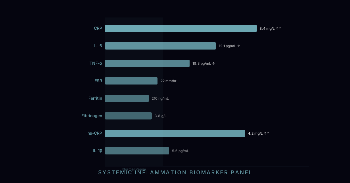
For inflammatory assessment in at-risk patients: Galectin-3 paired with the full complement of inflammation markers — hsCRP, fibrinogen, homocysteine, Lp-PLA2 (vascular inflammation), oxidized LDL, and myeloperoxidase (MPO, macrophage activation) — provides a comprehensive picture of inflammatory phenotype that can guide targeted interventions.
The future of galectin-3 monitoring likely includes serial testing every 6 to 12 months in patients receiving interventions targeting fibrosis, with the expectation that falling galectin-3 — like falling NT-proBNP during heart failure treatment — will serve as a biomarker-guided treatment response endpoint. This treat-to-target approach, well established for LDL cholesterol and blood pressure, represents the emerging standard for fibrosis management in high-risk patients.