Normal Female Testosterone: Ranges, Sources, and Why It Matters
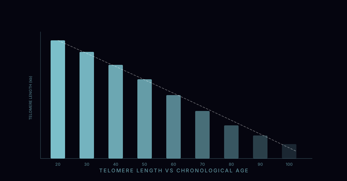
Testosterone is produced throughout a woman's reproductive life, peaking in the mid-20s and declining gradually thereafter — not in the abrupt fashion of estrogen at menopause, but in a steady decade-by-decade attrition that accelerates with ovarian aging and adrenal aging. A woman at 40 has approximately 50% of the testosterone she had at 20. The three production sources each contribute differently across the lifespan: the ovaries produce testosterone via theca cells stimulated by LH; the adrenal cortex produces testosterone and its precursors DHEA and DHEA-S; and peripheral tissues — adipose, skin, and muscle — convert adrenal androgens via 5-alpha reductase and aromatase into active androgens and estrogens.
Normal total testosterone in women is 15–70 ng/dL. Free testosterone — the biologically active fraction not bound to sex hormone binding globulin (SHBG) or albumin — is 0.3–1.9 pg/mL. These are the values that matter for symptom assessment, and they require attention to measurement methodology: most immunoassay-based testosterone tests used in routine labs have poor precision at female-range concentrations and should be confirmed with liquid chromatography-tandem mass spectrometry (LC-MS/MS) when clinical decisions are being made.
Why Low Testosterone in Women Is Chronically Under-Diagnosed
Despite decades of basic science demonstrating that androgen receptors are expressed in female brain, bone, muscle, skin, and cardiovascular tissue — and that testosterone plays distinct physiological roles in women — the clinical recognition of androgen insufficiency in women has lagged far behind. Several factors contribute to this diagnostic gap.
First, no FDA-approved testosterone product exists with a specific indication for women in the United States. Without a regulatory framework defining normal ranges and therapeutic targets in women, there has been less commercial pressure to generate the large-scale trial data that would support formal diagnostic criteria. Second, the symptoms of low testosterone in women (fatigue, low libido, reduced motivation, muscle loss) overlap substantially with those of low estrogen, low thyroid, iron deficiency, and depression — making attribution to androgen deficiency specifically difficult in clinical practice. Third, many physicians are simply unaware that women require testosterone, or are reluctant to prescribe an off-label treatment.
The result is a clinical situation in which a perimenopausal woman complaining of fatigue that has not resolved with estrogen therapy, low libido unresponsive to psychotherapy, and difficulty building muscle despite consistent resistance training may go undiagnosed for years because testosterone was never measured — or was measured with an immunoassay insufficiently sensitive to detect clinically relevant differences at female-range concentrations.
SHBG: The Hidden Variable in Testosterone Status
Sex hormone binding globulin (SHBG) is a hepatic glycoprotein that binds testosterone with high affinity, rendering it biologically inactive. Only free testosterone — and, to a lesser extent, albumin-bound testosterone — can enter cells and activate androgen receptors. SHBG is therefore as important as total testosterone in determining androgen status, and elevated SHBG is one of the most common causes of functional testosterone deficiency in women who have apparently normal total testosterone levels.
SHBG is raised by: oral estrogen (oral HRT, combined oral contraceptives — this is a crucial and frequently overlooked clinical point: women starting oral estrogen therapy often experience reduced libido and energy not because estrogen is causing this, but because oral estrogen raises SHBG, sharply reducing free testosterone), oral thyroid hormone at higher doses, certain anticonvulsants, and insulin sensitivity (low SHBG correlates with insulin resistance and polycystic ovary syndrome, where total testosterone is often normal or high but free testosterone is elevated). SHBG is lowered by: androgens themselves, obesity, insulin resistance, high protein intake, and hypothyroidism.
The practical implication: a woman prescribed oral estradiol (as part of HRT) who reports that her libido has worsened since starting therapy should have her SHBG checked. If SHBG is significantly elevated (above 100–120 nmol/L), switching from oral to transdermal estrogen (which does not undergo first-pass hepatic metabolism and does not raise SHBG) will often restore free testosterone without requiring testosterone supplementation.
Testing Protocol: How to Get Meaningful Results
A useful testosterone evaluation in women requires at minimum: total testosterone, free testosterone (or calculated free testosterone derived from total testosterone and SHBG), and SHBG. Additional context from DHEA-S (the major adrenal androgen precursor), LH, FSH, estradiol, and thyroid function is often valuable to identify the source of reduced androgen production and to rule out other contributors to the symptom complex.
Timing matters. Testosterone in premenopausal women peaks in the early-to-mid follicular phase (approximately days 3–10 of the cycle), is slightly lower in the luteal phase, and follows a diurnal pattern peaking in the early morning. Blood draws should ideally be performed in the morning (7–9am) and in the early follicular phase. Testing during the luteal phase will yield values that may be 10–20% lower than follicular-phase values, potentially misclassifying a borderline-normal result as deficient.
Method selection is critical. Standard immunoassay testosterone tests used in most commercial labs (LabCorp, Quest standard panels) are not adequately sensitive for female-range testosterone measurement. Equilibrium dialysis free testosterone or LC-MS/MS total testosterone should be specifically requested when evaluating women for androgen insufficiency. Some laboratories offer "female hormone panels" with LC-MS/MS methodology — these are substantially more informative than routine testosterone immunoassays at female-range concentrations.
Testosterone After Menopause: A Gradual Decline with Specific Consequences
Unlike estrogen, which falls precipitously at menopause, testosterone declines more gradually — roughly 50% per decade from peak — but the menopausal transition does accelerate the decline as ovarian production decreases. Women who undergo surgical menopause (bilateral oophorectomy) experience a more acute testosterone reduction of approximately 40–50% immediately post-surgery, since the ovaries contribute substantially to testosterone production even after menopause.
The clinical consequences of post-menopausal androgen decline most clearly established in RCT evidence include: hypoactive sexual desire disorder (HSDD), reduced sexual satisfaction, and diminished sexual responsiveness. The 2019 Global Consensus Position Statement — published jointly in multiple endocrinology journals — stated that there is Level 1 evidence (from randomised placebo-controlled trials) supporting testosterone therapy for postmenopausal women with HSDD, and that the benefits outweigh the risks of physiological-dose therapy. Beyond HSDD, the evidence for testosterone's effects on bone density, lean mass, cognitive function, and mood in postmenopausal women is accumulating but not yet at Level 1 — observational data and smaller RCTs are consistent in showing benefit, but confirmatory large randomised trials are still underway.
Topical Testosterone Therapy: What Is Available and How It Works
In the United States, no testosterone product is FDA-approved specifically for women, but off-label prescribing of male testosterone products at fractionated doses is an established clinical practice supported by published evidence. Common approaches include:
- AndroGel 1.62% (men's formulation): prescribed at a fraction of the male dose — typically 0.5 mg/day (approximately 1/10th the lowest male dose) applied to the inner forearm or inner thigh.
- Compounded testosterone cream or gel: prepared by a compounding pharmacy, typically at 0.5–1 mg/day for systemic effects or 2–4 mg for topical vulvovaginal use for local tissue effects.
- AndroFeme 1% (Australia): the only approved female testosterone product globally; a 1% testosterone cream applying 0.5 mg/day, with clinical trial data demonstrating efficacy and safety for HSDD in postmenopausal women.
Transdermal delivery is preferred over injectable testosterone for women because it provides stable, physiological concentrations without the supraphysiologic peaks that follow intramuscular injection at male doses. Testosterone pellet implants are also used by some practitioners, though the dose precision and reversibility of pellets is inferior to topical preparations.
Bone Density, Cardiovascular Health, and Longevity
Testosterone has direct effects on bone through androgen receptors expressed in osteoblasts and osteoclasts — independent of its aromatization to estrogen. Observational studies in postmenopausal women consistently show positive correlations between endogenous testosterone levels and bone mineral density at hip and spine. RCT data from testosterone trials in postmenopausal women shows modest but significant improvements in bone density at the hip and lumbar spine over 2 years.
Cardiovascular effects at physiological female doses are generally favorable or neutral: testosterone does not raise LDL, does not adversely affect blood pressure, and may modestly reduce visceral fat accumulation (which is independently associated with cardiovascular risk). The concern that testosterone would virilize cardiovascular risk profiles — as it does at supraphysiologic male doses with anabolic misuse — has not been borne out at female physiological dosing in the existing trial evidence.
The longevity angle is less directly studied but mechanistically compelling: maintaining muscle mass, bone density, metabolic rate, and cognitive function — all of which testosterone supports in women — are independently associated with healthy aging and reduced all-cause mortality. Whether testosterone supplementation in women translates to longevity outcomes specifically will require long-term trials that have not yet been completed.